 Copyright © Michael Richmond.
This work is licensed under a Creative Commons License.
Copyright © Michael Richmond.
This work is licensed under a Creative Commons License.
Let's take a look at how a scintillation detector works. How does it convert a gamma ray into an electrical pulse that can be counted?
When a gamma ray approaches an atom, there are several things that can happen.

One can use conservation of energy and momentum to derive formulae for the energy of the gamma-ray and the electron after an episode of Compton scattering:
Note the general result: the gamma ray always has less energy than it started, but there is an upper limit to the amount of energy it can lose. That means that the energy of the ejected electron may range from zero to a maximum value, which is again less than the energy of the incoming gamma ray.
Let us concentrate on possibilities 2 and 3. We'll begin with photoelectric absorption. The energy of the freed electron must be
In our lab, some typical values are
so it should be clear that the energy given to the liberated electron is almost exactly the same as the energy of the incident gamma ray, to a tiny fraction of a percent.
So, at this point, we have one electron with a boatload of kinetic energy. How can we turn this into a signal we can detect? The answer is to convert the electron's kinetic energy into visible light. The mechanism involves many collisions with atoms in a special sort of crystal: sodium iodide, doped with a small amount of thallium to replace a small fraction (0.1% to 0.4%) of the sodium atoms. The function of the thallium is to modify the electronic properties of the crystal. In an pure sodium iodide crystal, one finds the usual situation: a valence band and a conduction band, separated by a large gap, in this case about 5.9 eV.
If an electron were excited into the conduction band, then managed to drop back down into the valence band by emitting energy as a photon, the wavelength of that photon would be around 210 nm, in the near-UV. Such a photon would immediately be absorbed by other atoms in the crystal, which would prevent it from leaving the crystal and being measured.
When a small amount of thallium is added to the crystal, new bands are created at a somewhat lower energies above the valence band. The term "exciton" is sometimes used to describe electrons in this state.
An electron excited into the conduction band may now drop down to the lowest energy in the conduction band (as usual), but now it may drop further by small steps into the exciton states. As the electron makes its way back to the valence band by these small steps, it may emit a photon with a lower energy and longer wavelength.

The lower energy and longer wavelength of this photon are important for the detection process in two ways:
          initial gamma ray  E   =   662,000 eV      for Cs-137

               x 10 percent      =    66,000 eV

               / 3 ev/photon     =    22,000 photons
So, each gamma ray may cause tens of thousands of
visible-light photons to be emitted from the crystal.
Remember that only a fraction of these photons will
go in the right direction -- towards the photomultiplier tube.
We find that gamma = 2.3 and so the initial velocity of the electron must be around v = 0.9 c, very close to the speed of light. At relativistic speeds, it takes only around 10-18 seconds to travel from one atom in a crystal to another. Even if the electron must travel many atoms between interactions, it can make tens of thousands of collisions in a very short time, perhaps 10-13 seconds.
The incoming visible photon knocks just one electron from the photocathode. That single electron is accelerated through a high voltage, causing it to strike a metal surface (a "dynode") at high speed. It knocks several electrons free, and they are in turn accelerated towards a second metal surface. This process is repeated a number of times, increasing the number of electrons at each stage. By the end of the photomultiplier tube, the single electron has turned into a cloud of many thousands of electrons, a signal which can easily be measured.
The size of each packet of charge striking the final dynode (the anode) in a photomultiplier tube indicates the number of photons which struck the photocathode; that, in turn, indicates the energy of the electron which was bumping into atoms in the crystal; that, in turn, indicates the energy of the original gamma ray. There are, after all, quite a few steps in the process:
So, when we look at the spectrum of a typical gamma-ray source, such as Cs-137, what should we expect to see?
If we had a PERFECT instrument, we might expect to see a spectrum like this:

since Cs-137 produces gamma rays with energies of 32.06, 36.40 and 661.7 keV.
But what we ACTUALLY see looks quite different:
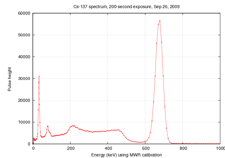
What has happened?
This broadening places a limit on the energy resolution of the instrument.
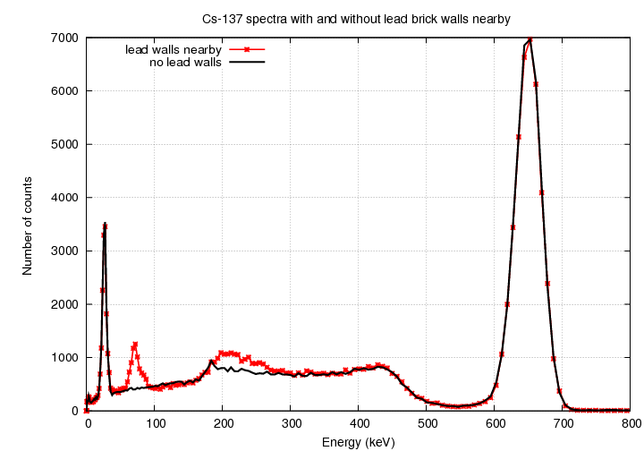
We usually place the radioactive source in the middle of a little structure made of lead bricks, to prevent radiation from escaping into the room at large. Some of the gamma rays from the source interact with the lead atoms in the bricks, creating lower-energy scattered gamma rays and also X-rays with the characteristic energy of the L-shell-to-K-shell transition in lead. The interactions with the lead shielding lead to several noticeable features in the spectrum. I removed the lead bricks and placed the source on an empty cardboard box so that it was at the same distance from the source as usual. Look at the difference in the spectrum:
Copyright © Michael Richmond.
This work is licensed under a Creative Commons License.